Neurobiologia del trattamento del PTSD e della depressione maggiore: cosa ci dicono le neuroimmagini
Titolo Rivista GRUPPI
Autori/Curatori Marco Pagani
Anno di pubblicazione 2017 Fascicolo 2016/1 Lingua Italiano
Numero pagine 9 P. 169-177 Dimensione file 158 KB
DOI 10.3280/GRU2016-001014
Il DOI è il codice a barre della proprietà intellettuale: per saperne di più
clicca qui
Qui sotto puoi vedere in anteprima la prima pagina di questo articolo.
Se questo articolo ti interessa, lo puoi acquistare (e scaricare in formato pdf) seguendo le facili indicazioni per acquistare il download credit. Acquista Download Credits per scaricare questo Articolo in formato PDF

FrancoAngeli è membro della Publishers International Linking Association, Inc (PILA)associazione indipendente e non profit per facilitare (attraverso i servizi tecnologici implementati da CrossRef.org) l’accesso degli studiosi ai contenuti digitali nelle pubblicazioni professionali e scientifiche
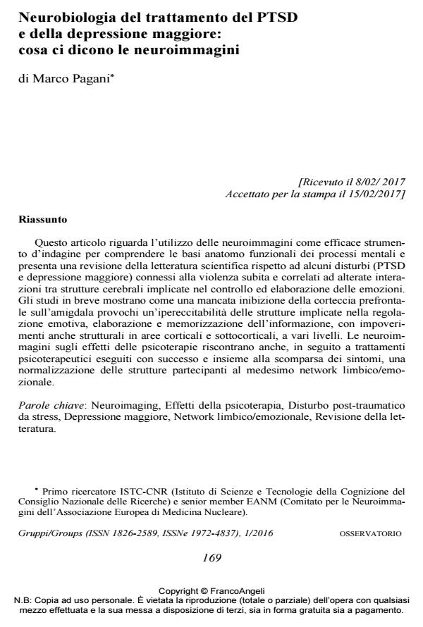
Questo articolo riguarda l’utilizzo delle neuroimmagini come efficace strumento d’indagine per comprendere le basi anatomo funzionali dei processi mentali e presenta una revisione della letteratura scientifica rispetto ad alcuni disturbi (PTSD e depressione maggiore) connessi alla violenza subita e correlati ad alterate interazioni tra strutture cerebrali implicate nel controllo ed elaborazione delle emozioni. Gli studi in breve mostrano come una mancata inibizione della corteccia prefrontale sull’amigdala provochi un’ipereccitabilità delle strutture implicate nella regolazione emotiva, elaborazione e memorizzazione dell’informazione, con impoverimenti anche strutturali in aree corticali e sottocorticali, a vari livelli. Le neuroimmagini sugli effetti delle psicoterapie riscontrano anche, in seguito a trattamenti psicoterapeutici eseguiti con successo e insieme alla scomparsa dei sintomi, una normalizzazione delle strutture partecipanti al medesimo network limbico/emozionale.;
Keywords:Neuroimaging, Effetti della psicoterapia, Disturbo post-traumatico da stress, Depressione maggiore, Network limbico/emozionale, Revisione della letteratura.
- Buchheim A., Viviani R., Kessler H., Kächele H., Cierpka M., Roth G., George C., Kernberg O.F., Bruns G. & Taubner S. (2012). Changes in Prefrontal-Limbic Function in Major Depression after 15 Months of Long-Term Psychotherapy. PLoS One, 1 March, 7, 3: e33745.
- Choi J., Jeong B., Polcari A., Rohan M.L. & Teicher M.H. (2012). Reduced Fractional Anisotropy in the Visual Limbic Pathway of Young Adults Witnessing Domestic Violence in Childhood. NeuroImage 59, 2: 1071-1079.
- Cisler J.M., Steele S.J., Smitherman S., Lenow J.K. & Kilts C.D. (2014). Neural Processing Correlate of Assaultive Violence Exposure and PTSD Symptoms during Implicit Threat Processing: A Network-level Analysis among Adolescent Girls. Psychiatry Research: Neuroimaging, 214: 238-246.
- de Greck M., Bölter A.F., Lehmann L., Ulrich C., Stockum E., Enzi B., Hoffmann T., Tempelmann C., Beutel M., Frommer J. & Northoff G. (2013). Changes in Brain Activity of Somatoform Disorder Patients during Emotional Empathy after Multimodal Psychodynamic Psychotherapy. Front Hum Neurosci., Aug., 16, 7: 410.
- Felmingham K., Kemp A., Williams L., Das P., Hughes G., Peduto A. & Bryant R. (2007). Changes in Anterior Cingulate and Amygdala after Cognitive Behavior Therapy of Posttraumatic Stress Disorder. Psychol. Sci., 18, 2: 127-129.
- Francati V., Vermetten E. & Bremner J.D. (2007). Functional Neuroimaging Studies in Posttraumatic Stress Disorder: Review of Current Methods and Findings. Depression and Anxiety, 24, 3: 202-218.
- Herringa R.J., Birna R.M., Ruttle P.L., Burghy C.A., Stodola D.E., Davidson R.J. & Essex M.J. (2013). Childhood Maltreatment is Associated with Altered Fear Circuitry and Increased Internalizing Symptoms by Late Adolescence. PNAS, November 19, 110, 47: 19119-19124.
- Kluetsch R.C., Ros T., Theberge J., Frewen P.A., Calhoun V.D., Schmahl C., Jetly R. & Lanius R.A. (2014). Plastic Modulation of PTSD Restingstate Networks and Subjective Wellbeing by EEG Neurofeedback. Acta Psychiatr. Scand., 130, 2: 123-136.
- Lansing K., Amen D.G., Hanks C. & Rudy L. (2005). High-resolution Brain SPECT Imaging and Eye Movement Desensitization and Reprocessing in Police Officers with PTSD. J. Neuropsychiatry Clin. Neurosci. 17, 4: 526-532.
- Lindauer R.J.L., Booij J., Habraken J.B.A., van Meijel E.P.M., Uylings H.B.M., Olff M., Carlier I.V.E., den Heeten G.J., van Eck Smit B.L.F. & Gersons B.P.R. (2008). Effects of Psychotherapy on Regional Cerebral Blood Flow during Trauma Imagery in Patients with Post-traumatic Stress Disorder: a Randomized Clinical Trial. Psychol. Med., 38, 04: 543-554. DOI: 10.1017/S0033291707001432
- Lindauer R.J.L., Vlieger E.J., Jalink M., Olff M., Carlier I.V.E., Majoie C.B.L.M., Den Heeten G.J. & Gersons B.P.R. (2005). Effects of Psychotherapy on Hippocampal Volume in Out-patients with Post-traumatic Stress Disorder: a MRI Investigation. Psychol. Med., 35, 10: 1421-1431. DOI: 10.1017/S003329170500524
- Nardo D., Högberg G., Looi J., Larsson S.A., Hällström T. & Pagani M. (2010). Grey Matter Changes in Posterior Cingulate and Limbic Cortex in PTSD are Associated with Trauma Load and EMDR Outcome. J. Psychiatry Research, 44: 477-485.
- Osuch E.A., Willis M.W., Bluhm R., Ursano R.J. & Drevets W.C. (2008). Neurophysiological Responses to Traumatic Reminders in the Acute Aftermath of Serious Motor Vehicle Collisions Using [15O]-H2O Positron Emission Tomography. Biol. Psychiatry, 64, 4: 327-335.
- Pagani M., Di Lorenzo G., Monaco L., Niolu C., Siracusano A., Verardo A.R., Lauretti G., Fernandez I., Nicolais G., Cogolo P. & Ammaniti M. (2011). Pretreat-ment, Intratreatment, and Posttreatment EEG Imaging of EMDR: Methodology and Preliminary Results from a Single Case. J. EMDR Pract. Res. 5, 2: 42-56. DOI: 10.1891/1933-3196.5.2.4
- Pagani M., Di Lorenzo G., Monaco L., Daverio A., Giannoudas I., La Porta P., Verardo A.R., Niolu C., Fernandez I. & Siracusano A. (2015). Neurobiological Response to EMDR Therapy in Clients with Different Psychological Traumas. Frontiers in Psychology – Psychology for Clinical Settings October, 6, Article 1614.
- Pagani M., Di Lorenzo G., Verardo A.R., Nicolais G., Monaco L., Lauretti G., Russo R., Niolu C., Ammaniti M., Fernandez I. & Siracusano A. (2012). Neurobiological Correlates of EMDR Monitoring – an EEG Study. PLoS One 7, 9: e45753.
- Pagani M., Hogberg G., Fernandez I. & Siracusano A. (2013). Correlates of EMDR Therapy in Functional and Structural Neuroimaging: a Critical Summary of Recent Findings. J. EMDR Pract. Res. 2013; 7, 1: 29-38. DOI: 10.1891/1933-3196.7.1.2
- Pagani M., Hogberg G., Salmaso D., Nardo D., Sundin O., Jonsson C., Soares J., Aberg Wistedt A., Jacobsson H., Larsson S.A. & Hallstrom T. (2007). Effects of EMDR Psychotherapy on 99mTc-HMPAO Distribution in Occupation-related Post-traumatic Stress Disorder. Nucl. Med. Commun., 28, 10: 757-765.
- Peres J.F.P., Foerster B., Santana L.G., Fereira M.D., Nasello A.G., Savoia M., Moreira Almeida A. & Lederman H. (2011). Police Officers under Attack: Resilience Implications of an fMRI Study. J. Psychiatr. Res., 45, 6: 727-734.
- Peres J.F.P., Newberg A.B., Mercante J.P., Simao M., Albuquerque V.E., Peres M.J.P. & Nasello A.G. (2007). Cerebral Blood Flow Changes during Retrieval of Traumatic Memories before and after Psychotherapy: a SPECT Study. Psychol. Med., 37, 10: 1481-1491. DOI: 10.1017/S003329170700997
- Rabe S., Zoellner T., Beauducel A., Maercker A. & Karl A. (2008). Changes in Brain Electrical Activity after Cognitive Behavioral Therapy for Posttraumatic Stress Disorder in Patients Injured in Motor Vehicle Accidents. Psychosom. Med., 70, 1: 13-19.
- Roy M.J., Francis J., Friedlander J., Banks-Williams L., Lande R.G., Taylor P., Blair J., McLellan J., Law W., Tarpley V., Patt I., Yu H., Mallinger A., Difede J., Rizzo A. & Rothbaum B. (2010). Improvement in Cerebral Function with Treatment of Posttraumatic Stress Disorder. Ann. N. Y. Acad. Sci., 1208: 142-149.
- Shin L.M., Orr S.P., Carson M.A., Rauch S.L., Macklin M.L., Lasko N.B., Peters P.M., Metzger L.J., Dougherty D.D., Cannistraro P.A., Alpert N.M., Fischman A.J. & Pitman R.K. (2004). Regional Cerebral Blood Flow in the Amygdala and Medial Prefrontal Cortex during Traumatic Imagery in Male and Female Vietnam Veterans with PTSD. Arch. Gen. Psychiatry, 61: 168-176.
- Siegel A., Bhatt S., Bhatt R. & Zalcman S. (2007). The Neurobiological Bases for Development of Pharmacological Treatments of Aggressive Disorders. Current Neuropharmacology, 2007, 5, 2: 135-147. DOI: 10.2174/15701590778086692
- Tomoda A., Sheu Y., Rabi K., Suzuki H., Navalta C.P., Polcari A. & Teicher M.H. (2011). Exposure to Parental Verbal Abuse is Associated with Increased Gray Matter Volume in Superior Temporal Gyrus. NeuroImage, 54: S280-S286.
- Wiswede D., Taubner S., Buchheim A., Munte1 T.F., Stasch M., Cierpka M., Kächele H., Roth G., Erhard P. & Kessler H. (2014). Tracking Functional Brain Changes in Patients with Depression under Psychodynamic Psychotherapy Using Individualized Stimuli. PLoS One, Oct., 9, 10: e109037.
- Bremner J.D. (2007). Functional Neuroimaging in Post-traumatic Stress Disorder. Expert Review of Neurotherapeutics, 7, 4: 393-405. DOI: 10.1586/14737175.7.4.39
- Brom D., Kleber R.J. & Defares P.B. (1989). Brief Psychotherapy for Post-traumat-ic Stress Disorders. J. Consult. Clin. Psychol., Oct., 57, 5: 607-12. DOI: 10.1037/0022-006X.57.5.60
- Bryant R.A., Felmingham K., Kemp A., Das P., Hughes G., Peduto A. & Williams L. (2008). Amygdala and Ventral Anterior Cingulate Activation Predicts Treatment Response to Cognitive Behaviour Therapy for Post-traumatic Stress Disorder. Psychol. Med., 38, 04: 555-561. DOI: 10.1017/S003329170700223
Marco Pagani, Neurobiologia del trattamento del PTSD e della depressione maggiore: cosa ci dicono le neuroimmagini in "GRUPPI" 1/2016, pp 169-177, DOI: 10.3280/GRU2016-001014