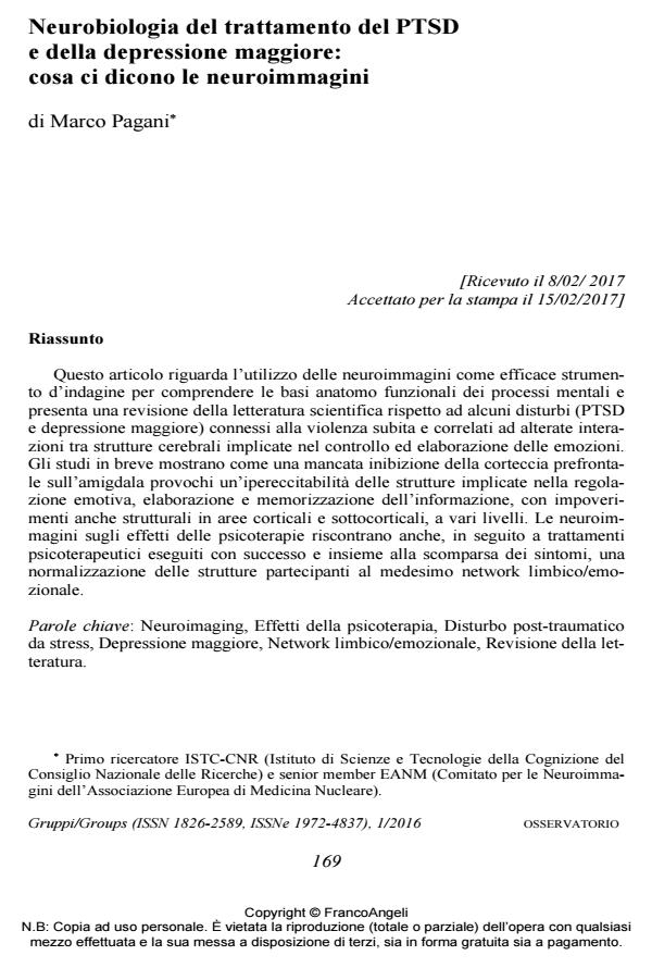
Neurobiologia del trattamento del PTSD e della depressione maggiore: cosa ci dicono le neuroimmagini
Journal title GRUPPI
Author/s Marco Pagani
Publishing Year 2017 Issue 2016/1
Language Italian Pages 9 P. 169-177 File size 158 KB
DOI 10.3280/GRU2016-001014
DOI is like a bar code for intellectual property: to have more infomation
click here
Below, you can see the article first page
If you want to buy this article in PDF format, you can do it, following the instructions to buy download credits

FrancoAngeli is member of Publishers International Linking Association, Inc (PILA), a not-for-profit association which run the CrossRef service enabling links to and from online scholarly content.
Neuroimaging is an efficient tool to investigate the anatomical and functional basis of mental processes. A revision of the scientific literature regarding conditions (PTSD and major depression) connected to violence and traumatic experiences is presented, exploring the connection between these syndromes and a modified interaction between cerebral structures implicated in the control and processing of emotions. Research shows how a missing inhibition of the prefrontal cortex on the amygdala leads to hyperexcitability of the structures involved in emotional regulation, elaboration and memorization of information and with impoverishments, some of which structural, in cortical and subcortical areas at multiple levels. Neu-roimaging on the effects of psychotherapy show, in successful psychotherapeutic treatments resulting in the disappearance of the symptoms, a normalisation of the structures regarding the same limbic emotional network. Neuroimaging is an efficient tool to investigate the anatomical and functional basis of mental processes. A revision of the scientific literature regarding conditions (PTSD and major depression) connected to violence and traumatic experiences is presented, exploring the connection between these syndromes and a modified interaction between cerebral structures implicated in the control and processing of emotions. Research shows how a missing inhibition of the prefrontal cortex on the amygdala leads to hyperexcitability of the structures involved in emotional regula-tion, elaboration and memorization of information and with impoverishments, some of which structural, in cortical and subcortical areas at multiple levels. Neuroimaging on the effects of psychotherapy show, in successful psychotherapeutic treatments resulting in the disappearance of the symptoms, a normalisation of the structures regarding the same limbic emotional network.
Keywords: Neuroimaging, Psychotherapy effects, Posttraumatic stress disorder (PTSD), Major depression, Limbic emotional network, Literature review
- Bremner J.D. (2007). Functional Neuroimaging in Post-traumatic Stress Disorder. Expert Review of Neurotherapeutics, 7, 4: 393-405. DOI: 10.1586/14737175.7.4.39
- Brom D., Kleber R.J. & Defares P.B. (1989). Brief Psychotherapy for Post-traumat-ic Stress Disorders. J. Consult. Clin. Psychol., Oct., 57, 5: 607-12. DOI: 10.1037/0022-006X.57.5.60
- Bryant R.A., Felmingham K., Kemp A., Das P., Hughes G., Peduto A. & Williams L. (2008). Amygdala and Ventral Anterior Cingulate Activation Predicts Treatment Response to Cognitive Behaviour Therapy for Post-traumatic Stress Disorder. Psychol. Med., 38, 04: 555-561. DOI: 10.1017/S003329170700223
- Buchheim A., Viviani R., Kessler H., Kächele H., Cierpka M., Roth G., George C., Kernberg O.F., Bruns G. & Taubner S. (2012). Changes in Prefrontal-Limbic Function in Major Depression after 15 Months of Long-Term Psychotherapy. PLoS One, 1 March, 7, 3: e33745.
- Choi J., Jeong B., Polcari A., Rohan M.L. & Teicher M.H. (2012). Reduced Fractional Anisotropy in the Visual Limbic Pathway of Young Adults Witnessing Domestic Violence in Childhood. NeuroImage 59, 2: 1071-1079.
- Cisler J.M., Steele S.J., Smitherman S., Lenow J.K. & Kilts C.D. (2014). Neural Processing Correlate of Assaultive Violence Exposure and PTSD Symptoms during Implicit Threat Processing: A Network-level Analysis among Adolescent Girls. Psychiatry Research: Neuroimaging, 214: 238-246.
- de Greck M., Bölter A.F., Lehmann L., Ulrich C., Stockum E., Enzi B., Hoffmann T., Tempelmann C., Beutel M., Frommer J. & Northoff G. (2013). Changes in Brain Activity of Somatoform Disorder Patients during Emotional Empathy after Multimodal Psychodynamic Psychotherapy. Front Hum Neurosci., Aug., 16, 7: 410.
- Felmingham K., Kemp A., Williams L., Das P., Hughes G., Peduto A. & Bryant R. (2007). Changes in Anterior Cingulate and Amygdala after Cognitive Behavior Therapy of Posttraumatic Stress Disorder. Psychol. Sci., 18, 2: 127-129.
- Francati V., Vermetten E. & Bremner J.D. (2007). Functional Neuroimaging Studies in Posttraumatic Stress Disorder: Review of Current Methods and Findings. Depression and Anxiety, 24, 3: 202-218.
- Herringa R.J., Birna R.M., Ruttle P.L., Burghy C.A., Stodola D.E., Davidson R.J. & Essex M.J. (2013). Childhood Maltreatment is Associated with Altered Fear Circuitry and Increased Internalizing Symptoms by Late Adolescence. PNAS, November 19, 110, 47: 19119-19124.
- Kluetsch R.C., Ros T., Theberge J., Frewen P.A., Calhoun V.D., Schmahl C., Jetly R. & Lanius R.A. (2014). Plastic Modulation of PTSD Restingstate Networks and Subjective Wellbeing by EEG Neurofeedback. Acta Psychiatr. Scand., 130, 2: 123-136.
- Lansing K., Amen D.G., Hanks C. & Rudy L. (2005). High-resolution Brain SPECT Imaging and Eye Movement Desensitization and Reprocessing in Police Officers with PTSD. J. Neuropsychiatry Clin. Neurosci. 17, 4: 526-532.
- Lindauer R.J.L., Booij J., Habraken J.B.A., van Meijel E.P.M., Uylings H.B.M., Olff M., Carlier I.V.E., den Heeten G.J., van Eck Smit B.L.F. & Gersons B.P.R. (2008). Effects of Psychotherapy on Regional Cerebral Blood Flow during Trauma Imagery in Patients with Post-traumatic Stress Disorder: a Randomized Clinical Trial. Psychol. Med., 38, 04: 543-554. DOI: 10.1017/S0033291707001432
- Lindauer R.J.L., Vlieger E.J., Jalink M., Olff M., Carlier I.V.E., Majoie C.B.L.M., Den Heeten G.J. & Gersons B.P.R. (2005). Effects of Psychotherapy on Hippocampal Volume in Out-patients with Post-traumatic Stress Disorder: a MRI Investigation. Psychol. Med., 35, 10: 1421-1431. DOI: 10.1017/S003329170500524
- Nardo D., Högberg G., Looi J., Larsson S.A., Hällström T. & Pagani M. (2010). Grey Matter Changes in Posterior Cingulate and Limbic Cortex in PTSD are Associated with Trauma Load and EMDR Outcome. J. Psychiatry Research, 44: 477-485.
- Osuch E.A., Willis M.W., Bluhm R., Ursano R.J. & Drevets W.C. (2008). Neurophysiological Responses to Traumatic Reminders in the Acute Aftermath of Serious Motor Vehicle Collisions Using [15O]-H2O Positron Emission Tomography. Biol. Psychiatry, 64, 4: 327-335.
- Pagani M., Di Lorenzo G., Monaco L., Niolu C., Siracusano A., Verardo A.R., Lauretti G., Fernandez I., Nicolais G., Cogolo P. & Ammaniti M. (2011). Pretreat-ment, Intratreatment, and Posttreatment EEG Imaging of EMDR: Methodology and Preliminary Results from a Single Case. J. EMDR Pract. Res. 5, 2: 42-56. DOI: 10.1891/1933-3196.5.2.4
- Pagani M., Di Lorenzo G., Monaco L., Daverio A., Giannoudas I., La Porta P., Verardo A.R., Niolu C., Fernandez I. & Siracusano A. (2015). Neurobiological Response to EMDR Therapy in Clients with Different Psychological Traumas. Frontiers in Psychology – Psychology for Clinical Settings October, 6, Article 1614.
- Pagani M., Di Lorenzo G., Verardo A.R., Nicolais G., Monaco L., Lauretti G., Russo R., Niolu C., Ammaniti M., Fernandez I. & Siracusano A. (2012). Neurobiological Correlates of EMDR Monitoring – an EEG Study. PLoS One 7, 9: e45753.
- Pagani M., Hogberg G., Fernandez I. & Siracusano A. (2013). Correlates of EMDR Therapy in Functional and Structural Neuroimaging: a Critical Summary of Recent Findings. J. EMDR Pract. Res. 2013; 7, 1: 29-38. DOI: 10.1891/1933-3196.7.1.2
- Pagani M., Hogberg G., Salmaso D., Nardo D., Sundin O., Jonsson C., Soares J., Aberg Wistedt A., Jacobsson H., Larsson S.A. & Hallstrom T. (2007). Effects of EMDR Psychotherapy on 99mTc-HMPAO Distribution in Occupation-related Post-traumatic Stress Disorder. Nucl. Med. Commun., 28, 10: 757-765.
- Peres J.F.P., Foerster B., Santana L.G., Fereira M.D., Nasello A.G., Savoia M., Moreira Almeida A. & Lederman H. (2011). Police Officers under Attack: Resilience Implications of an fMRI Study. J. Psychiatr. Res., 45, 6: 727-734.
- Peres J.F.P., Newberg A.B., Mercante J.P., Simao M., Albuquerque V.E., Peres M.J.P. & Nasello A.G. (2007). Cerebral Blood Flow Changes during Retrieval of Traumatic Memories before and after Psychotherapy: a SPECT Study. Psychol. Med., 37, 10: 1481-1491. DOI: 10.1017/S003329170700997
- Rabe S., Zoellner T., Beauducel A., Maercker A. & Karl A. (2008). Changes in Brain Electrical Activity after Cognitive Behavioral Therapy for Posttraumatic Stress Disorder in Patients Injured in Motor Vehicle Accidents. Psychosom. Med., 70, 1: 13-19.
- Roy M.J., Francis J., Friedlander J., Banks-Williams L., Lande R.G., Taylor P., Blair J., McLellan J., Law W., Tarpley V., Patt I., Yu H., Mallinger A., Difede J., Rizzo A. & Rothbaum B. (2010). Improvement in Cerebral Function with Treatment of Posttraumatic Stress Disorder. Ann. N. Y. Acad. Sci., 1208: 142-149.
- Shin L.M., Orr S.P., Carson M.A., Rauch S.L., Macklin M.L., Lasko N.B., Peters P.M., Metzger L.J., Dougherty D.D., Cannistraro P.A., Alpert N.M., Fischman A.J. & Pitman R.K. (2004). Regional Cerebral Blood Flow in the Amygdala and Medial Prefrontal Cortex during Traumatic Imagery in Male and Female Vietnam Veterans with PTSD. Arch. Gen. Psychiatry, 61: 168-176.
- Siegel A., Bhatt S., Bhatt R. & Zalcman S. (2007). The Neurobiological Bases for Development of Pharmacological Treatments of Aggressive Disorders. Current Neuropharmacology, 2007, 5, 2: 135-147. DOI: 10.2174/15701590778086692
- Tomoda A., Sheu Y., Rabi K., Suzuki H., Navalta C.P., Polcari A. & Teicher M.H. (2011). Exposure to Parental Verbal Abuse is Associated with Increased Gray Matter Volume in Superior Temporal Gyrus. NeuroImage, 54: S280-S286.
- Wiswede D., Taubner S., Buchheim A., Munte1 T.F., Stasch M., Cierpka M., Kächele H., Roth G., Erhard P. & Kessler H. (2014). Tracking Functional Brain Changes in Patients with Depression under Psychodynamic Psychotherapy Using Individualized Stimuli. PLoS One, Oct., 9, 10: e109037.
Marco Pagani, Neurobiologia del trattamento del PTSD e della depressione maggiore: cosa ci dicono le neuroimmagini in "GRUPPI" 1/2016, pp 169-177, DOI: 10.3280/GRU2016-001014