Correlati neurobiologici nell’anziano con malattia di alzheimer e altre demenze

Titolo Rivista RICERCHE DI PSICOLOGIA
Autori/Curatori Antonio Guaita
Anno di pubblicazione 2013 Fascicolo 2012/2-3
Lingua Italiano Numero pagine 18 P. 389-406 Dimensione file 224 KB
DOI 10.3280/RIP2012-002016
Il DOI è il codice a barre della proprietà intellettuale: per saperne di più clicca qui

Qui sotto puoi vedere in anteprima la prima pagina di questo articolo.

Se questo articolo ti interessa, lo puoi acquistare (e scaricare in formato pdf) seguendo le facili indicazioni per acquistare il download credit. Acquista Download Credits per scaricare questo Articolo in formato PDF
Anteprima articolo

FrancoAngeli è membro della Publishers International Linking Association, Inc (PILA)associazione indipendente e non profit per facilitare (attraverso i servizi tecnologici implementati da CrossRef.org) l’accesso degli studiosi ai contenuti digitali nelle pubblicazioni professionali e scientifiche

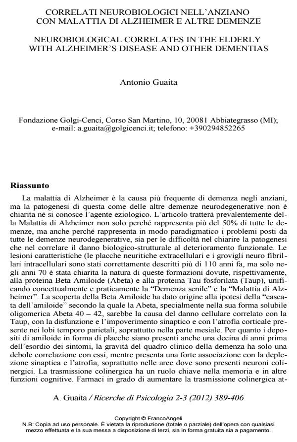
La malattia di Alzheimer e la causa piu frequente di demenza negli anziani, ma la patogenesi di questa come delle altre demenze neurodegenerative non e chiarita ne si conosce l’agente eziologico. L’articolo trattera prevalentemente della Malattia di Alzheimer non solo perche rappresenta piu del 50% di tutte le demenze, ma anche perche rappresenta in modo paradigmatico i problemi posti da tutte le demenze neurodegenerative, sia per le difficolta nel chiarire la patogenesi che nel correlare il danno biologico-strutturale al deterioramento funzionale. Le lesioni caratteristiche (le placche neuritiche extracellulari e i grovigli neuro fibrillari intracellulari sono stati correttamente descritti piu di 110 anni fa, ma solo negli anni 70 e stata chiarita la natura di queste formazioni dovute, rispettivamente, alla proteina Beta Amiloide (Abeta) e alla proteina Tau fosforilata (Taup), unificando concettualmente e praticamente la "Demenza senile" e la "Malattia di Alzheimer". La scoperta della Beta Amiloide ha dato origine alla ipotesi della "cascata dell’amiloide" secondo la quale la Abeta, specialmente nella sua forma solubile oligomerica Abeta 40 - 42, sarebbe la causa del danno cellulare correlato con la Taup, con la disfunzione e l’impoverimento sinaptico e con l’atrofia corticale presente nei lobi temporo parietali, soprattutto nella parte mesiale. Per quanto i depositi di amiloide in forma di placche siano presenti anche una decina di anni prima dell’esordio dei sintomi, la gravita del quadro clinico della demenza ha solo una debole correlazione con essi, mentre presenta una forte associazione con la deplezione sinaptica e l’atrofia, soprattutto nelle aree dove sono presenti neuroni colinergici. La trasmissione colinergica ha un ruolo chiave nella memoria e in altre funzioni cognitive. Farmaci in grado di aumentare la trasmissione colinergica at- traverso l’inibizione della acetilcolinesterasi sono oggi in uso come trattamento sintomatico del deficit cognitivo. Oggi la neuro tossicita viene quindi ascritta alle forme solubili di Beta Amiloide, mentre le placche potrebbero essere interpretate anche come conseguenza, quasi un meccanismo di inattivazione e di difesa dalla tossicita oligomerica. Il danno vascolare puo di per se provocare demenza, ma ha un forte ruolo anche nella malattia di Alzheimer, dove pure sono sempre presenti anche danni vascolari di diversa gravita tanto che le due entita non sono spesso distinguibili. Altri tipi di demenza sono associati con la degenerazione Lobare Frontotemporale o con la deposizione di corpi di Lewy, ma in eta avanzata questi quadri non sono piu cosi distinti e le sovrapposizioni sono molto frequenti, sia cliniche che neuropatologiche, anche con il quadro neuro anatomico di persone senza demenza. Questo pone alla definizione dei correlati neurobiologici delle demenze degenerative problemi non solo tecnici ma anche di natura concettuale ed epistemologica.;

Keywords:Demenza, malattia di Alzheimer, neuropatologia, beta amiloide, marcatori biologici.

  1. Marcello, E., Epis R., Saraceno, C., & Di Luca, M. (2012). Synaptic dysfunction in Alzheimer’s disease. Advances in Experimental Medicine and Biology, 970, 573
  2. Bateman, R.J., Xiong, C., Benzinger, T.L., Fagan, A.M., Goate, A., Fox, N.C., Marcus, D.S., Cairns, N.J., Xie, X., Blazey, T.M., Holtzman, D.M., Santacruz, A., Buckles, V., Oliver, A., Moulder, K., Aisen, P.S., Ghetti, B., Klunk, W.E., McDade, E., Martins, R.N., Masters, C.L., Mayeux, R., Ringman, J.M., Rossor, M.N., Schofield, P.R., Sperling, R.A., Salloway, S., Morris, J.C., & Dominantly Inherited Alzheimer Network (2012). Clinical and biomarker changes in dominantly inherited Alzheimer’s disease. The New England Journal of Medicine, 367, 795-804
  3. Bennett, D.A., Schneider, J.A., Bienias, J.L., Evans, D.A., & Wilson R.S. (2005). Mild cognitive impairment is related to Alzheimer disease pathology and cerebral infarctions. Neurology, 64, 834-841.
  4. Birks, J. (2006). Cholinesterase inhibitors for Alzheimer’s disease. Cochrane Database Syst Rev, CD005593
  5. Bowen, D.M., Smith, C.B., White, P., & Davison, A.N. (1976). Neurotransmitterrelated enzymes and indices of hypoxia in senile dementia and other abiotrophies. Brain, 99, 459-496
  6. Braak, H., & Braak, E. (1991). Neuropathological stageing of Alzheimer-related changes. Acta Neuropathologica, 82, 239-259
  7. Braak, H., & Del Tredici, K. (2011). Alzheimer’s pathogenesis, is there neuron- to-neuron propagation? Acta Neuropathologica, 121, 589-595.
  8. Chui, H.C., Zarow, C., Mack, W.J., Ellis, W.G., Zheng, L., & Jagust, W.J. (2006). Cognitive impact of subcortical vascular and Alzheimer’s disease pathology. Annals of Neurology, 60, 677-687
  9. Coleman, P.D., & Yao, P.J. (2003). Synaptic slaughter in Alzheimer’s disease. Neurobiology of Aging, 24, 1023-1027
  10. DeKosky, S.T, Scheff, S.W., & Styren, S.D. (1996). Structural correlates of cognition in dementia, quantification and assessment of synapse change. Neurodegeneration, 5, 417-421
  11. Duff, K., Eckman, C., Zehr, C., Yu, X., Prada, C.M., Perez-tur, J., Hutton, M., Buee, L., Harigaya, Y., Yager, D., Morgan, D., Gordon, M.N., Holcomb, L., Refolo, L., Zenk, B., Hardy, J., & Younkin, S. (1996). Increased amyloid- beta42(43) in brains of mice expressing mutant presenilin 1. Nature, 383, 710-713.
  12. Farina, E., Baglio, F., Caffarra, P., Magnani, G., Scarpini, E., Appollonio, I., Bascelli, C., Cheldi, A., Nemni, R., Franceschi, M., Italian Group for Lewy Body Dementia and Dementias Associated to Parkinsonism, Messa, G., Mantovani, F., Bellotti, M., Olivotto, F., Alberoni, M., Isella, V., Regazzoni, R., Schiatti, E,. Vismara, C., Falautano, M., Barbieri, A., Restelli, I., Fetoni, V., Donato, M., Zuffi, M., & Castiglioni S. (2009). Frequency and clinical features of Lewy body dementia in Italian memory clinics. Acta Biomedica, 80, 57-64.
  13. Fotuhi, M., Hachinski, V., & Whitehouse, P.J. (2009). Changing perspectives regarding late-life dementia. Nature Reviews Neurology, 5, 649-658.
  14. Giannakopoulos, P., Herrmann, F.R., Bussière, T., Bouras C., Kövari E., Perl D. P., Morrison J.H., Gold G., & Hof, P.R. (2003). Tangle and neuron numbers, but not amyloid load, predict cognitive status in Alzheimer’s disease. Neurology, 60, 1495-1500.
  15. Glenner, G.G., & Wong, C.W. (1984). Alzheimer’s disease and Down’s syndrome, sharing of a unique cerebrovascular amyloid fibril protein. Biochemical and Biophysical Research Communications, 122, 1131-1135
  16. Gong, Y., Chang, L., Viola, K.L., Lambert, M.P., Frinch, C.E., Krafft, G.A., & Klein, W.L. (2003). Alzheimer’s disease-affected brain, presence of oligomeric A ligands (ADDLs) suggests a molecular basis for reversibile memory loss. Proceedings of the National Academy of Sciences of the United States of America, 100, 10417-10422.
  17. Grundke-Iqbal, I., Iqbal, K., Tung, Y.C., Quinlan, M., Wisniewski, H.M., & Binder, L.I. (1986). Abnormal phosphorylation of the microtubule-associated protein tau (tau) in Alzheimer cytoskeletal pathology. Proceedings of the National Academy of Sciences of the United States of America, 83, 4913–4917
  18. Hardy, J., & Selkoe, D.J. (2002). The amyloid hypothesis of Alzheimer’s disease, progress and problems on the road to therapeutics. Science, 297, 353-356
  19. Holtzman, D.M. (2011). CSF biomarkers for Alzheimer’s disease, current utility and potential future use. Neurobiol Aging, 32 Suppl 1, S4-9.
  20. Hoover, B.R., Reed, M.N., Su, J., Penrod, R.D., Kotilinek, L.A., Grant, M.K., Pitstick, R., Carlson, G.A., Lanier, L.M., Yuan, L.L., Ashe, K.H., & Liao, D. (2010). Tau mislocalization to dendritic spines mediates synaptic dysfunction independently of neurodegeneration. Neuron, 68, 6, 1067-1081
  21. Hyman, B.T., Phelps, C.H., Beach, T.G., Bigio, E.H., Cairns, N.J., Carrillo, M.C., Dickson, D.W., Duyckaerts, C., Frosch, M.P., Masliah, E., Mirra, S.S., Nelson, P.T., Schneider, J.A., Thal, D.R., Thies, B., Trojanowski, J.Q., Vinters, H.V., & Montine, T.J. (2012). National Institute on Aging-Alzheimer’s Association guidelines for the neuropathologic assessment of Alzheimer’s disease. Alzheimers Dement, 8, 1-13.
  22. Hyman, B.T., & Trojanowski, J.Q. (1997). Consensus recommendations for the postmortem diagnosis of Alzheimer disease from the National Institute on Aging and the Reagan Institute Working Group on diagnostic criteria for the neuropathological assessment of Alzheimer disease. Journal of Neuropathology & Experimental Neurology, 56, 1095–1097
  23. Katzman, R. (1976). Editorial, The prevalence and malignancy of Alzheimer disease. A major killer. Archives of Neurology,33, 217-218
  24. Kessels, H. W., Nguyen, L.N., Nabavi, S., & Malinow, R. (2010). The prion protein as a receptor for amyloid-b. Nature 466, E3-E5.
  25. Knopman, D.S., DeKosky, S.T., Cummings, J.L., Chui, H., Corey-Bloom, J., Relkin, N., Small, G.W., Miller, B., & Stevens, J.C. (2001). Practice parameter, diagnosis of dementia (an evidence-based review). Report of the Quality Standards Subcommittee of the American Academy of Neurology. Neurology, 56, 1143-1153.
  26. Koh, J.Y., Yang, L.L., & Cotman, C.W. (1990). β-amyloid protein increases the vulnerability of cultured cortical neurons to excitotoxic damage. Brain Research, 533, 315-320
  27. Lee, H.G., Perry, G., Moreira, P.I., Garrett, M.R., Liu, Q., & Zhu, X.W. (2005). Tau phosphorylation in Alzheimer’s disease, pathogen or protector? Trends in Molecular Medicine, 11, 164-169
  28. Mandybur, T.I. (1975). The incidence of cerebral amyloid angiopathy in Alzheimer’s disease. Neurology,25, 120-126
  29. Roth, M., Tomlinson, B.E., & Blessed, G. (1967). The relationship between quantitative measures of dementia and of degenerative changes in the cerebral grey matter of elderly subjects. Proc R Soc Med., 60, 254-60
  30. Schliebs, R., & Arendt, T. (2011). The cholinergic system in aging and neuronal degeneration. Behavioural Brain Research, 221, 555-563.
  31. Sherrington, R., Rogaev, E.I., Liang, Y., Rogaeva, E.A., Levesque, G., Ikeda, M., Chi, H., Lin, C., Li, G., Holman, K., Tsuda, T., Mar, L., Foncin, J.F., Bruni, A.C., Montesi, M.P., Sorbi, S., Rainero, I., Pinessi, L., Nee, L., Chumakov, I., Pollen, D., Brookes, A., Sanseau, P., Polinsky, R.J., Wasco, W., Da Silva, H. A., Haines, J.L., Perkicak-Vance, M.A., Tanzi, R.E., Roses, A.D., Fraser, P. E., Rommens, J.M., & St George-Hyslop, P.H. (1995). Cloning of a gene bearing missense mutations in early-onset familial Alzheimer’s disease. Nature, 375, 754-760.
  32. Sonnen, J.A., Santa Cruz, K., Hemmy, L.S., Woltjer, R., Leverenz, J.B., Montine, K.S., Jack, C.R., Kaye J., Lim, K., Larson, E.B., White, L., & Montine, T.J. (2011). Ecology of the aging human brain. Archives of Neurology, 68, 1049- 1056
  33. Summers, W.K., Majovski, L.V., Marsh, G.M., Tachiki, K., & Kling, A. (1986). Oral tetrahydroaminoacridine in long-term treatment of senile dementia, Alzheimer type. The New England Journal of Medicine, 315, 1241-1245.
  34. Tanzi, R.E., Moir, R.D., & Wagner, S.L. (2004). Clearance of Alzheimer’s Abeta peptide, the many roads to perdition. Neuron., 43, 605-608
  35. Terry, R.D., Masliah, E., & Salmon, D.P. (1991). Physical basis of cognitive alterations in Alzheimer’s disease, synapse loss is the major correlate of cognitive impairment. Annals of Neurology, 30, 572-580.
  36. Vossel, K.A., Zhang, K., Brodbeck, J., Daub, A.C., Sharma, P., Finkbeiner, S., Cui, B., & Mucke, L. (2010). Tau reduction prevents Abeta-induced defects in axonal transport. Science, 330, 198.
  37. Walsh, D.M., Klyubin, I., Fadeeva, J.V., Kulln, W.K., Anwyl, R., Wolfe, M.S, Rowan, M.J., & Selkoe, D.J. (2002). Naturally secreted oligomers of amyloid beta protein potently inhibit hippocampal long-term potentiation in vivo. Nature, 416, 535-539.
  38. Wei, W., Nguyen, L.N., Kessels, H.W., Hagiwara, H., Sisodia, S., & Malinow, R. (2010). Amyloid b from axons and dendrites reduces local spine number and plasticity. Nature Neuroscience, 13, 190-196
  39. Whitehouse, P.J., Price, D.L., Struble, R.G., Clark, A.W., Coyle, J.T., & Delon, M.R. (1982). Alzheimer’s disease and senile dementia, loss of neurons in the basal forebrain. Science, 215, 1237-1239
  40. Wilson, R.S., Leurgans, S.E., Boyle, P.A., Schneider, J.A., & Bennett, D.A. (2010). Neurodegenerative basis of age-related cognitive decline. Neurology, 75, 1070-1078
  41. Yiannopoulou, K.G., & Papageorgiou, S.G. (2013). Current and future treatments for Alzheimer’s disease. Therapeutic Advances in Neurological Disorders, 6, 19-33.
  42. Zhang, Y., He J.S., Wang, X., Wang, J., Bao, F.X., Pang, S.Y., Yin, F., Hu, H.G., Peng, X.L., Sun, W.M., Zheng, Y.P., Hou, L.L., & Hong, T. (2011). Administration of amyloid-β42 oligomer-specific monoclonal antibody improved memory performance in SAMP8 mice. Journal of Alzheimer's Disease, 23, 551- 561
  43. Zlokovic, B. V. (2004). Clearing amyloid through the blood-brain barrier. Journal of Neurochemistry, 89, 807-811.
  44. Zlokovic, B.V. (2005). Neurovascular mechanisms of Alzheimer’s neurodegeneration. Trends in Neuroscience, 28, 202-208.
  45. Riedel-Heller, S.G., Busse, A., Aurich, C., Matschinger, H., & Angermeyer, M. C. (2001). Incidence of dementia according to DSM-III-R and ICD-10, results of the Leipzig Longitudinal Study of the Aged (LEILA75+), Part 2. The British Journal of Psychiatry, 179, 255-260
  46. Ravaglia, G., Forti, P., Maioli, F., Sacchetti, L., Mariani, E., Nativio, V., Talerico, T., Vettori, C., & Macini, P.L. (2002). Education, occupation, and prevalence of dementia, findings from the Conselice study. Dementia and Geriatric Cognitive Disorders, 14, 90-100.
  47. Ratnavalli, E., Brayne, C., Dawson, K., & Hodges, J. R. (2002). The prevalence of frontotemporal dementia. Neurology, 58, 1615-1621
  48. Querfurth, H.W., & La Ferla, F.M. (2010). Alzheimer’s disease. The New England Journal of Medicine, 36, 329-344.
  49. Puzzo, D., Vitolo, O., Trinchese, F., Jacob, J.P., Palmeri, A., & Arancio, O. (2005). Amyloid-beta peptide inhibits activation of the nitric oxide/cGMP/cAMP-responsive element-binding protein pathway during hippocampal synaptic plasticity. Journal of Neuroscience, 25, 6887-6897.
  50. Neto, M.R. (2002). Volumetric MRI measurements can differentiate Alzheimer’s disease, mild cognitive impairment, and normal aging. International Psychogeriatrics, 14, 59-72.
  51. Nelson, P.T., Schmitt, F.A., Lin, Y., Abner, E.L., Jicha, G.A., Patel, E., Thomason, P.C., Neltner, J.H., Smith, C.D., Santacruz, K.S., Sonnen, J.A., Poon, L.W., Gearing, M., Green, R.C., Woodard, J.L., Van Eldik, L.J., & Kryscio, R.J. (2011). Hippocampal sclerosis in advanced age, clinical and pathological features. Brain, 134, 1506-1518
  52. Nagy, Z., Yilmazer-Hanke, D.M., Braak, H., Braak, E., Schultz, C., & Hanke, J. (1998). Assessment of the pathological stages of Alzheimer’s disease in thin paraffin sections, a comparative study. Dementia and Geriatric Cognitive Disorders, 9, 140-144
  53. Montine, T.J., Phelps, C.H., Beach, T.G., Bigio, E.H., Cairns, N.J., Dickson, D.W., Duyckaerts, C., Frosch, M.P., Masliah, E., Mirra, S.S., Nelson, P.T., Schneider, J.A., Thal, D.R., Trojanowski, J.Q., Vinters, H.V., & Hyman, B.T. (2012). National Institute on Aging-Alzheimer’s Association guidelines for the neuropathologic assessment of Alzheimer’s disease, a practical approach. Acta Neuropathologica, 123, 1-11.
  54. Montine, T.J., & Larson, E.B. (2009). Late-life dementias, does this unyielding global challenge require a broader view? Jama, 302, 2593-2594
  55. Mondragón-Rodríguez, S., Basurto-Islas, G., Lee, H.G., Perry, G., Zhu, X., Castellani, R.J., & Smith, M.A. (2010). Causes versus effects, the increasing complexities of Alzheimer’s disease pathogenesis. Expert Review of Neurotherapeutics, 10, 683-691
  56. Mirra, S.S., Heyman, A., McKeel, D., Sumi, S.M., Crain, B.J., Brownlee, L.M., Vogel, F.S., Hughes, J.P., van Belle, G., & Berg, L. (1991). The Consortium to Establish a Registry for Alzheimer’s Disease (CERAD). Part II. Standardization of the neuropathologic assessment of Alzheimer’s disease. Neurology,41, 479-486
  57. Meda, L., Cassatella, M.A., Szendrei, G.I., Otvo, L.Jr., Baron, P., Villalba, M., Ferari, D., & Rossi, F. (1995). Activation of microglia cells by beta-amyloid protein and interferon-gamma. Nature, 374, 647-650
  58. Masters, C.L., Simms, G., Weinman, N.A., Multhaup, G., McDonald, B.L., & Beyreuther, K. (1985). Amyloid plaque core protein in Alzheimer disease and Down syndrome. Proceedings of the National Academy of Sciences of the United States of America, 82, 4245-4249

Antonio Guaita, Correlati neurobiologici nell’anziano con malattia di alzheimer e altre demenze in "RICERCHE DI PSICOLOGIA " 2-3/2012, pp 389-406, DOI: 10.3280/RIP2012-002016